
Spectroscopy Applications
Sequentially Timed All-optical Mapping Photography (STAMP) uses a Coherent Astrella to capture video bursts on timescales of ~100 fs to several ns.
This article shows how an ultra-stable femtosecond amplifier enables high data output in 2D IR spectroscopic studies of biological membranes and surfactants.

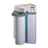
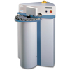
The OPIS 35™ laser accessory is a companion to the Hyperflux™ PRO Plus (HFPP) Raman Spectrometer.

This experiment demonstrates that chemometric modes can be easily transferred between the HyperFlux™ PRO Plus and the Process Guardian™ Raman spectrometers without extraordinary data manipulation.

Protecting consumer safety with Misa SERS analyser.

Identification of acids through a plastic squeeze container.
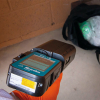
Handheld Raman system is capable of material identification in complex environments.
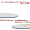
Mira spectrometers equipped with ORS capture a large interrogation area in a very short time, analysing all of the ingredients in a single scan.
In this application note, an XRF spectrometer is used to analyse several elements/oxides in ores in a matter of minutes.
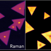
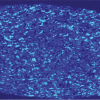
CVD-grown MoS2 flakes are analysed using Raman and PL imaging with a Raman microscope.
A wavelength dispersive X-ray fluorescence spectroscopy technique for iron ore analysis is described that minimises analysis time.
Enhancement of SERS features by electrochemical activation in non-aqueous media.
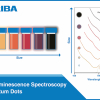
emission spectra, which is dependent on their size and composition.

A good portable Raman spectrometer bridges the gap in performance, capability and size between handheld and benchtop Raman spectroscopy.
Checking the content uniformity of tablets is a crucial step in quality control of these dosage forms—but it is slow and tedious. With IR laser imaging a comprehensive content uniformity is finally possible in less than 10 min.
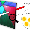
First, the LSPR of spherical colloidal AuNPs is investigated as a function of particle diameter. Then, the chemical sensing potential of AuNPs is demonstrated by tracking the LSPR during an induced aggregation experiment.

Two chemical analyses from one tablet using Raman to identify the major component and SERS for the minor component.

The catalogue showcases Hiden's solutions for analysing dissolved species in liquids.

A multiparameter determination within one minute using NIR spectroscopy.

Commercial SERS substrates are readily available, making SERS an accessible technique for low-concentration detection. In this application note, residual phosmet insecticide on apple skin is detected using SERS.
