Katie Ember
University of Edinburgh and University of Strathclyde. E-mail: [email protected]
Monday
On the walk to Haymarket station, I avoid the stares of curious commuters. I try not to be envious of their tiny handbags or compact briefcases. I attempt to maintain a steady course and a nonchalant look. Because at 8 o’clock in the morning, it’s hard to remain inconspicuous whilst wheeling a large yellow Pelican case through the streets of Edinburgh.
Pelican cases were originally designed for military and law enforcement use. They are durable, waterproof and typically used to transport “sensitive equipment”, e.g. cameras or guns, but my cargo is significantly nerdier: a 785 nm portable Raman spectrometer. After some trials at the University of Edinburgh, I’m now returning the device to its home at the University of Strathclyde in Glasgow.
I drag the case through the barriers, heave it from platform to train and spend the rest of the journey reading papers whilst remaining acutely aware of how much room the spectrometer is taking up in the luggage rack.
I’m halfway through the final year of my PhD in Optical Medical Imaging, and collaboration between universities and departments has been a large part of my project. The aim of my research is to develop a way of detecting liver damage using spectroscopy and it’s about as interdisciplinary as you can get. To achieve this, I’ve been working with physicists, chemists, biologists and transplant surgeons in both the cities of Edinburgh and Glasgow. It involves a lot of travel, but it’s been a great way to learn about a whole host of different subjects and there’s very rarely a dull day.
Fifty-eight minutes after the train pulls out of Haymarket, I arrive at Strathclyde’s Technology and Innovation centre—affectionately called the TIC. For such an unpleasant acronym, it’s a gorgeous building. From a bird’s eye view it is triangular, and its seven stories are home to state-of-the-art labs carrying out some of Scotland’s most innovative research, ranging from renewable energy to photonics.
Haymarket Station: Train station towards the West end of Edinburgh, lesser known than the more central Waverley Bridge station
The Meadows: central park in Edinburgh, beloved by runners, barbeque-ers and people doing strange acrobatic sports
The Royal Infirmary: one of Edinburgh’s multiple hospitals; relatively new and in the southern outskirts
King’s Buildings: the science campus of the University of Edinburgh
George Square: the University’s Central campus. A mix of Edwardian houses and brutalist concrete surrounding gardens open to students in the summer
My supervisor here is Karen Faulds, Professor of Pure and Applied Chemistry at the Centre for Nanotechnology. Professsor Faulds’ work focuses on the biological applications of Raman spectroscopy, a technique which uses the inelastic scattering of laser light to yield molecular information about samples.
There are a number of variations on the theme of Raman spectroscopy and Professor Faulds’ group are making use of a number of them. For example, surface-enhanced Raman spectroscopy (SERS) employs metal nanoparticles (or other metal surfaces) to enhance the otherwise weak Raman signal.
These particles and surfaces can be functionalised with molecules that bind to specific cells or react to particular environmental conditions, telling us about organ health. It is not a technique I’ve been using, but Faulds’ group have used SERS to detect multiple types of pathogenic bacteria.1,2
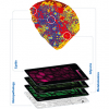
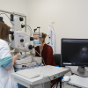
After catching up briefly with some of the other students at the TIC, I place the spectrometer carefully back on its lab bench and switch on the Leica multiphoton microscope. A scientist probably shouldn’t have favourite instruments as they all have their advantages and limitations, but it’s hard not to love the multiphoton.
Leica multiphoton microscope at the University of Strathclyde.
If someone said “imagine a fancy microscope designed by Batman”, that’s what the Leica looks like—a huge, gleaming, jet-black exterior housing a glimmering assortment of filters, mirrors and lenses. The software, however, seems to have been developed by the world’s biggest fan of rainbows and, in all honesty, it’s a welcome addition to the dark microscopy room.
A major advantage of the Leica is that it possesses multiple excitation sources, lending it CARS, SRS and fluorescence imaging capabilities. Stimulated Raman spectroscopy (SRS) and coherent anti-Stokes Raman spectroscopy (CARS) both make use of two lasers: a pump beam and a Stokes beam. By tuning these lasers to specific frequencies, you can enhance the signal at the Raman shift of interest. Although this limits the spectral information acquired, it is possible to gain high-quality, high-resolution images of your target peak in an extremely short time.
For example, you can label a molecule of interest with an alkyne tag. As alkynes have a Raman signal in the middle of the “cell silent” region, the distribution of the tagged molecule can be rapidly imaged.
After a day at the microscope, I take the train back to Haymarket carrying nothing but my small backpack and a USB full of images.
Tuesday
Thesis-writing day. My alarm goes off at 6:30 am: I’m much more productive in the mornings. Besides, it’s exam season and the library becomes packed with undergrads as the day goes on—so packed sometimes the only available desk space is in the downstairs cafe. My tiny purple laptop is on its last legs and the Edinburgh Uni library has hundreds of desktop computers, complete with software necessary to reference, create graphs and do statistical analysis. It’s not surprising that it’s so popular.
I’ve been writing my results into a paper and processing data as I go, which has been incredibly helpful. Paper writing has also ensured that I’ve been using multiple methods throughout my PhD to confirm my findings. Although Raman spectra are informative, no single experimental technique is sufficient to categorically prove something, especially within the field of biological sciences. For this reason, I’ve been using well-characterised techniques such as NMR spectroscopy and histological staining in parallel with Raman to support my findings.
NMR spectroscopy uses a magnetic field and radio-frequency electromagnetic radiation to induce energetic transitions in a sample. The peaks are sharper than those obtained with Raman or IR spectroscopy, so you can resolve individual molecules more easily. However, NMR requires samples in the form of biofluids or a biopsy which limits its application to the clinic. Nevertheless, I’ve used NMR to measure changes in the metabolic profile of damaged livers versus control livers and these are the spectra I’ve been focussing on today.
By 11:15 am, my eyes can’t take any more NMR spectra and I’m craving caffeine, so I leave my desk and walk to one of the tiny coffee boxes in George Square. It looks a bit like a TARDIS (it is in fact an old police box) and they make excellent Americanos.
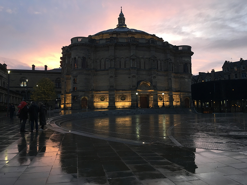
MacEwan Hall near George Square at sunset.
Back to the library, to an email saying that lab meeting is still going ahead. Lab meeting. Cool. I had absolutely not forgotten about that. My chemistry supervisor, Dr Colin Campbell, is currently on sabbatical in Colorado where he is developing ways of culturing and imaging cells in 3D, and we have lab meetings via Skype.
The rest of the team meet in Room 232 in the School of Chemistry and it’s a good chance to catch up with other members of the group. Many of us work in different departments, so it can be easy to forget that there are others who have faced similar problems at another point in their PhD. We have all benefitted from advice after presenting our work at these meetings.
Other students in Dr Campbell’s group have been using SERS in a similar way to researchers at Strathclyde, but functioning the nanoparticles with redox-sensitive molecules. As the redox state of the environment changes, so do the Raman spectra of the molecules attached to the metal nanoparticles. Therefore, the Raman spectra can be used as a readout for the redox state within 3D cell cultures or even tumour models.3
After the meeting, I go straight to the University gym. The building is only a few years old and is kitted out with cardio rooms, free weights, fixed weights, basketball and squash courts, and a climbing wall. I aim to exercise at least five times per week; whether that’s at the gym, playing football or running around the Meadows. It allows me to switch off completely from PhD life; much as I love science, it’s good to separate work and home.
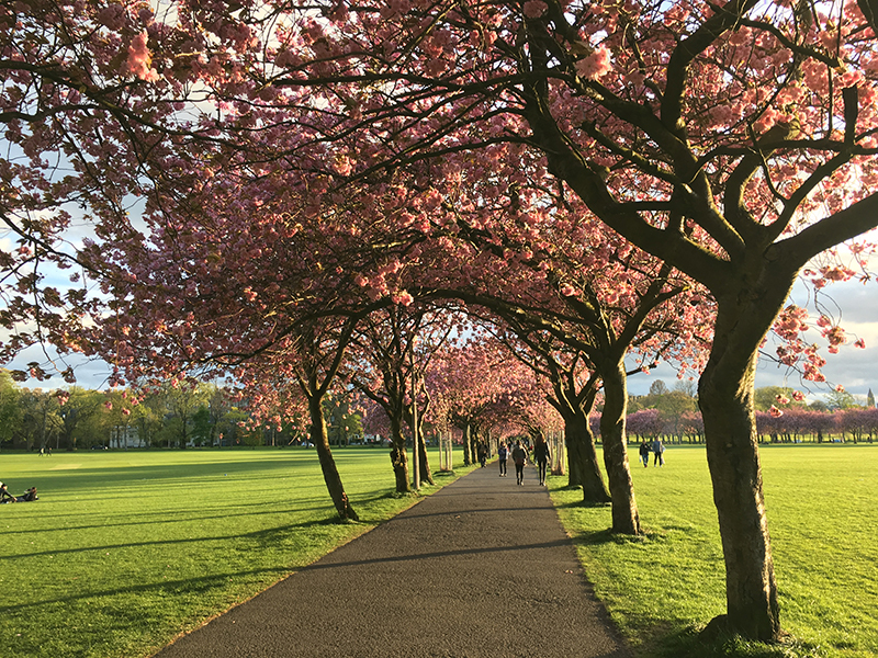
Spring in the Meadows near the George Square library.
Wednesday
The number 24 bus winds its way through what feels like the whole of Edinburgh, but finally we arrive at the Royal Infirmary. This morning, I have a meeting with Mr Gabriel Oniscu, a transplant surgeon I’m working with at the Royal. One of the reasons we’re developing a way of detecting liver damage using spectroscopy is to see if we can assess the health of donated livers.
Liver transplants take around six to eight hours and they’re remarkable to observe. The entire team—composed of nurses, surgeons and anaesthetists—remain alert and focused throughout the process, each carrying out their own critical role with utmost care. However, the one point where the transplant team use subjectivity is during assessment of the donated liver. The operating surgeon mostly achieves this by visually inspecting the liver and checking it isn’t fatty or visibly injured. Being able to detect damage at a molecular level in real time would be invaluable.
Unfortunately, introducing a laser-based device into the well-oiled transplant process will be far from simple. Mr Oniscu and transplant theatre sister Ms Fiona Hunt have been vital sources of practical, clinically-relevant information. Any spectroscopic solution must be able to take into account surgical lights, motion and variations in biochemistry from patient to patient. The device needs to give molecular information without requiring a surgeon to analyse raw spectral data. And it has to be able to do so rapidly.
After Mr Oniscu and I have talked through my most recent results, I join another PhD student who is having liver samples graded by a pathologist and has invited me along. To assess the samples, the pathologist is using a microscope with multiple eyepieces, so I and the other student can see exactly what he’s talking about. We’re learning a lot.
“These white spots are vacuoles, it’s sort of a mark for reversible cell damage… And here, you have necrosis, it’s only in a small area though.” Necrosis is a type of cell death that transplant surgeons want to avoid. The liver has a remarkable capacity to regenerate, but serious levels of necrosis can result in scarring, loss of function and cancer.
After our mini pathology tutorial, we grab a coffee from the hospital cafe, before heading back to the office to try to make sense of the new results.
Thursday
“Let’s try lower acquisition time, higher laser power; just to see what happens.”
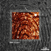
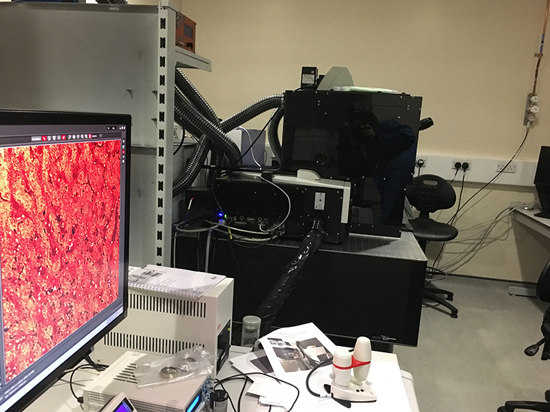
This morning, I’m teaching a new PhD student how to use the Renishaw InVia Raman spectrometer. It’s been more time-consuming than we had anticipated, especially as her samples are organoids (tiny spheres of cells) and we’re not sure what data to expect. With the Renishaw, there are so many parameters you can change: laser power, acquisition time, streamline or point acquisition, lens, map size... and there are two lasers—532 nm and 785 nm excitation.

Renishaw InVia at the Edinburgh Bioquarter.
Moreover, there are unique challenges to face when using biological samples. Sometimes you’ll be using frozen sections, other time cells growing in a monolayer, sometimes biofluids such as serum. Each sample will have different optical properties and hence the experimental set-up must be adjusted accordingly. It is also important to make sure that the surrounding medium isn’t giving a strong Raman signal that would drown out the signal of the molecules you are trying to detect.
And then there is the problem of laser intensity. When I first started using liver tissue, I set the spectrometer to run a mapping experiment for 8 hours. The idea was to take tens of thousands of spectra from a tiny rectangle within the tissue. I returned the next morning to find a perfect 500 µm by 700 µm oblong had been vaporised from my sample.
After we’ve determined the optimal parameters for the experiment, I take the bus to the Business School at George Square. I know what you’re thinking—where does studying Business fit into detecting liver damage? It’s a good question: I am a member of OPTIMA, the Centre for Doctoral Training in Optical Medical Imaging. Many scientists lack the skills to take their ideas from bench to business, so this four-year programme has been specifically designed to help PhD students gain training in entrepreneurship as well as interdisciplinary science.
In this module, our group has been learning about the steps required to take an implant from Phase I trials right the way through to market. We’ve learned about intellectual property, ethics and finance—it’s been a fascinating course and I wish all biomedical scientists were able to gain an insight into these processes.
Friday
Friday morning is the Forbes group lab meeting, based at the Centre for Regenerative Medicine (CRM). Professor Stuart Forbes is my clinical supervisor and a consultant hepatologist at the Royal Infirmary. His lab at the CRM specialises in liver regeneration—a complex and fascinating process. Given time, human livers are able to regain structure and function even after large sections are damaged or removed; and the Forbes group aim to discover the cellular mechanisms behind this process.4
The other lab members mostly use light microscopy, biochemical and proteomic techniques, but they’ve been endlessly patient in teaching me how to process tissue samples and interpret the data in terms of the biological state of the liver. The diversity of the knowledge and skills in the lab is astounding and they’ve given me access to cell lines, mouse models, rat models, patient biopsies and discarded livers. Cell lines are easier to obtain and less biologically complex than whole organs. However, they are not a true reflection of the state within living organisms so it is useful to be able to carry out experiments on tissue samples too.
After the meeting, I spend the afternoon working on a conference presentation from the Campbell office at the School of Chemistry. Every few months, I try to present at a conference or do some form of public engagement: the impact of research is limited if you don’t communicate it with a wider audience. Fortunately, my supervisors are all very encouraging of science communication and I’ve had the chance to present in London, Barcelona and Canada to audiences ranging from school children to surgeons.
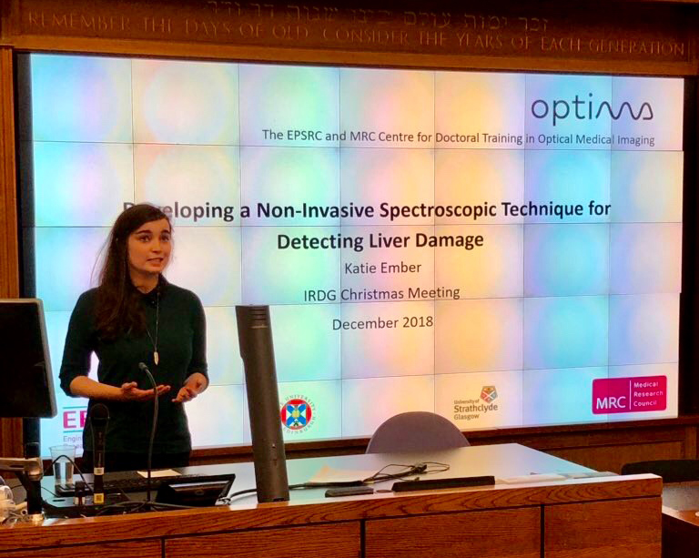
Presenting a talk at the IRDG Christmas meeting, December 2018.
Once the PowerPoint is in good shape, it’s time to head to The Southern—a pub patronised by PhDs and postdocs alike. It’s the weekend and our conversations turn to upcoming holidays, sports and our life plans for next year. Failed experiments and PhD problems are forgotten.
Concluding thoughts
A PhD isn’t for everyone and, before starting, it is worth researching the project, the lab culture and the city you’re going to call home. Postgraduate degrees can be psychological and emotional rollercoasters: I’ve had moments where NMR tubes have shattered, where experiments have run on for seventeen hours and my entire first year’s work has been filed under “optimisation”. But these have been insignificant setbacks in three incredible years.
I’ve been free to learn countless new research techniques, develop communication skills and explore multiple areas of science. All things considered; I wouldn’t change the last few years for anything.
References
- K. Gracie, D. Lindsay, D. Graham and K. Faulds, “Bacterial meningitis pathogens identified in clinical samples using a SERS DNA detection assay”, Anal. Methods 7, 1269–1272 (2015). https://doi.org/10.1039/C5AY00063G
- H. Kearns, R. Goodacre, L. Jamieson, D. Graham and K. Faulds, “SERS detection of multiple antimicrobial-resistant pathogens using nanosensors”, Anal. Chem. 89, 12666–12673 (2017). https://doi.org/10.1021/acs.analchem.7b02653
- L.E. Jamieson, A. Jaworska, J. Jiang, M. Baranska, D.J. Harrison and C.J. Campbell, “Simultaneous intracellular redox potential and pH measurements in live cells using SERS nanosensors”, Analyst 140, 2330–2335 (2015). https://doi.org/10.1039/C4AN02365J
- S.J. Forbes and P.N. Newsome, “Liver regeneration — mechanisms and models to clinical application”, Nat. Rev. Gastroenterol. Hepatol. 13(8), 473–485 (2016). https://doi.org/10.1038/nrgastro.2016.97