2 March 2021 to 5 March 2021
[email protected]
[email protected]
This is a fully interactive online event. The course provides an in-depth introduction to the world of time-resolved fluorescence microscopy with a focus on life science applications. The program combines lectures by experts in the field with practical sessions on systems from the market-leading companies Nikon, Olympus, Zeiss, and PicoQuant.
The course’s content is geared towards both novice researchers as well as those having some previous experience with time-resolved methods and applications. The lectures and practical sessions provide participants with a better understanding of the following topics and applications:
- Instrumentation and data analysis
- Correlation spectroscopy methods: FCS, FCCS, FLCS
- Imaging methods: FLIM, rapid FLIM, FRET, FLIM-FRET
- Single molecule microscopy

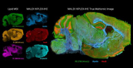
![Targeted proton transfer charge reduction (tPTCR) nano-DESI mass spectrometry imaging of liver tissue from orally dosed rat (Animal 3). a) optical image of a blood vessel within liver tissue. b) Composite ion image of charge-reduced haeme-bound α-globin (7+ and 6+ charge states; m/z 2259.9 and m/z 2636.3 respectively, red) and the charged-reduced [FABP+bezafibrate] complex (7+ and 6+ charge states; m/z 2097.5 and m/z 2446.9 respectively, blue). c) Ion image composed from charge-reduced haeme-bound α-globin (7+ and 6+ charge states) showing abundance in blood vessels. d) Ion image composed from charge-reduced [FABP+bezafibrate] complex (7+ and 6+ charge states) showing abundance in bulk tissue and absence in the blood vessel. Reproduced from https://doi.org/10.1002/ange.202202075 under a CC BY licence. Light and mass spectromert imaging of tissue samples](/sites/default/files/styles/see_also/public/news/MSI%20drug-protein%20complex-w.jpg?itok=K57jqSou)
