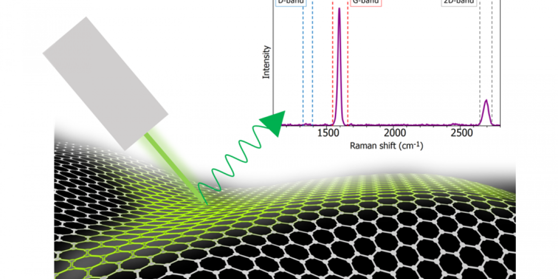
The structure and bonding of graphene make it well suited for Raman spectroscopy. Graphene is one atom thick carbon; the carbon atoms are arranged in a hexagonal lattice. The sp2 carbon bonds result in highly polarisable π bonds which give an intense Raman signal. This is because the vibrational mode is “Raman active” and this occurs when the molecular polarisability changes during the vibration. Raman spectroscopy can be used in the evaluation of graphene quality and thickness. Since Raman microscopy is non-destructive and has high spatial and spectral resolution, the technique is well-suited for gathering detailed information about the graphene film. There is also minimal or no sample preparation required for Raman spectroscopy, further adding to its ease of use and low damage risk. In this application note, an Edinburgh Instruments RM5 Raman Microscope is used to highlight how Raman microscopy an essential tool for any material scientist researching graphene.

